
Figure 3.
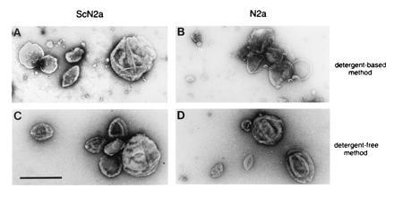
Ultrastructure of isolated CLDs. (A and B) CLDs were isolated by the ice-cold Triton X-100 detergent method. (C and D) CLDs isolated by the detergent-free procedure. Samples were prepared from ScN2a (A and C) and N2a cells (B and D). (Bar = 0.5 μm.)