FIG. 3.
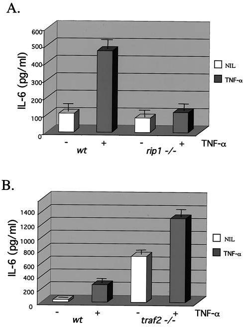
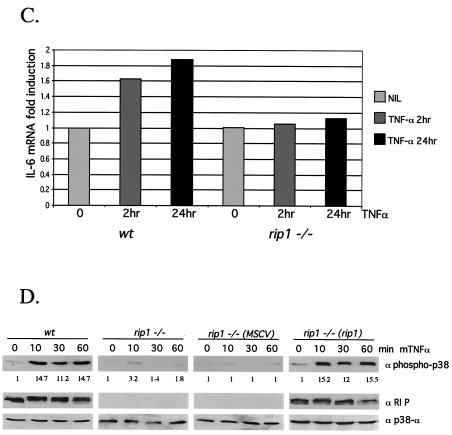
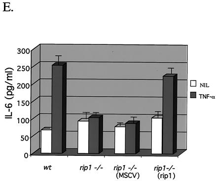
Decreased TNF-α-induced IL-6 production in rip1−/− cells. (A) Wild-type and rip1−/− MEF were plated at 3 × 10⁴ cells per well on 24-well plates and left untreated or treated with 10 ng of TNF-α/ml for 24 h. The supernatants were then analyzed for IL-6 levels with the OptEIA mouse IL-6 ELISA kit (PharMingen catalog no. 2653KI). (B) TNF-α-induced IL-6 production in traf2−/− cells. Wild-type and traf2−/− cells were plated on 24-well plates at 3 × 10⁴ cells per well and left untreated or treated with 10 ng of TNF-α/ml for 24 h. The supernatants were then analyzed for IL-6 levels with the OptEIA mouse IL-6 ELISA kit. The amount of IL-6 is presented as the mean ± standard deviation of triplicate observations. Similar data were obtained in six independent experiments with six independent rip−/− MEF lines and five traf2−/− MEF lines. (C) Decreased TNF-α-induced IL-6 mRNA in rip1−/− MEF. Wild-type and rip1−/− MEF were left untreated or treated with TNF-α (10 ng/ml) for 2 and 24 h. The amount of IL-6 mRNA and GAPDH mRNA was measured by RNase protection assay. The protected RNA was detected by autoradiography after denaturing polyacrylamide gel electrophoresis and was quantitated by PhosphorImager analysis. For clarity, exposure time varied for autoradiography for IL-6 and GAPDH mRNAs. Similar data were obtained in three separate experiments. (D) TNF-α-induced p38 MAPK activation in rip1−/− MEF infected with RIP1 retrovirus. rip1−/− MEF were infected with vector alone (MSCV) or with a RIP1 retrovirus [rip1−/− (rip1)]. Wild-type, rip1−/−, rip1−/− (MSCV), and rip1−/− (rip1) cells were left untreated or stimulated with TNF-α for the time periods indicated, and p38 MAPK activity was measured by immunoblotting with an anti-phospho-p38 Ab. The RIP1 and p38α expression was determined by immunoblotting with anti-RIP and anti-p38α Abs. The percentage of green fluorescent protein-positive cells in the infected populations was determined by flow cytometry. Results from one of three independent infection experiments are shown. (E) TNF-α-induced IL-6 production is increased in rip1−/− MEF infected with a RIP1 retrovirus. Wild-type, rip1−/−, and rip1−/− MEF infected with vector (MSCV) or with a RIP1 retrovirus [rip1−/− (rip1)] were plated at 3 × 10⁴ cells per well on 24-well plates and left untreated or treated with 10 ng of TNF-α/ml for 24 h. The supernatants were then analyzed for IL-6 levels with the OptEIA mouse IL-6 ELISA kit. The amount of IL-6 is presented as the mean ± standard deviation of triplicate observations.


