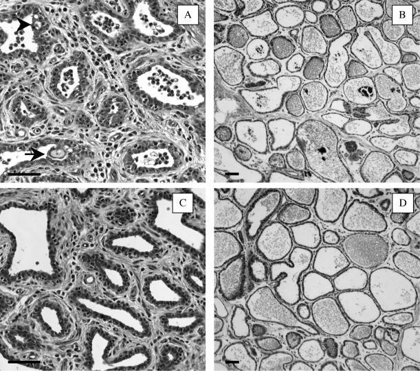
FIG. 1.
Photomicrographs of the gland cistern (A and C) and the mammary parenchymal (B and D) tissues dissected from SEC-inoculated (A and B) and PBS-inoculated (C and D) mammary glands. SEC (100 μg) or PBS alone was inoculated into lactating mammary glands, and tissues were collected at 28 h after inoculation. Each photomicrograph is typical of those for three experimental cows. The arrows indicate invaginations of the ductular epithelium. The arrowheads indicate cytoplasmic vacuolations of epithelial cells. Bars, 50 μm.

