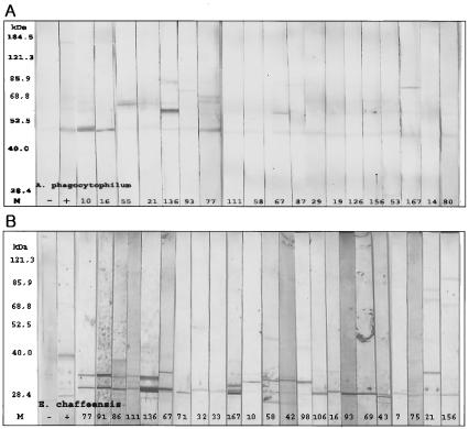
FIG. 3.
Western blot assay of electrophoretically separated gradient-purified A. phagocytophilum (A) and E. chaffeensis (B) antigens reacted with IFA-positive sera. Lanes: M, protein size marker; −, negative control; +, positive control; the lane numbers represent sample numbers. (A) Twenty-five serum specimens (83.3%) reacted with an A. phagocytophilum antigen of approximately 44 kDa presumed to be Msp2. The reaction patterns were divided into three groups: 11 serum specimens reacted only with the 44-kDa antigen, 8 reacted with the 44-kDa antigen and another antigen between 60 and 85 kDa, and 5 reacted with the 44-kDa antigen and two other antigens between 60 and 160 kDa. (B) Twenty-nine (74.4%) serum specimens reacted with purified E. chaffeensis antigens. The reaction patterns of 32 serum specimens were also divided into three groups: 15 reacted only with antigens of 28 kDa, 10 reacted with two antigens between 28 and 35 kDa, and 4 reacted with three or four antigens between 26 and 100 kDa.

