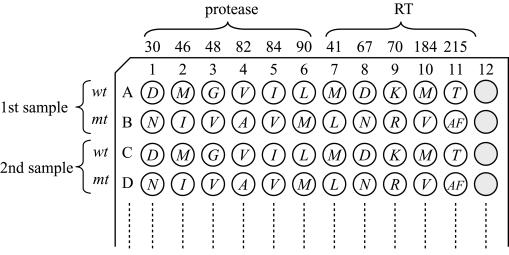
FIG. 3.
Alignment of hybridization probes in a 96-plate format. There are two rows for each sample. The first row of each sample is coated with wild-type (wt) detection probes, and the second row of each sample is coated with mutant (mt) probes. The italicized letter in each well demonstrates the detectable amino acid pattern.

