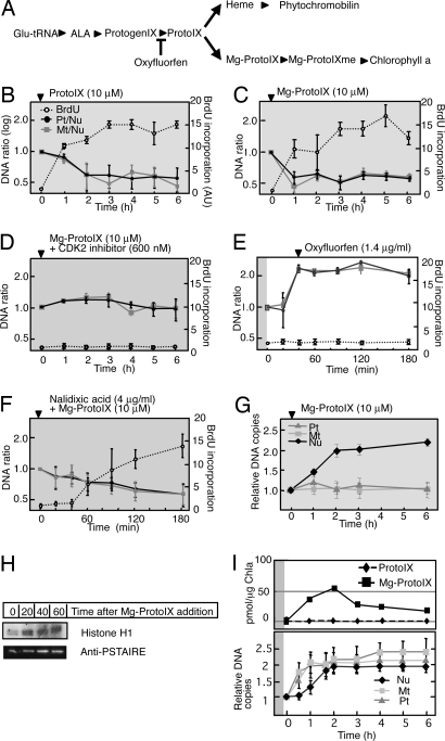
Fig. 2.
Effects of tetrapyrroles and an inhibitor of ProtoIX biosynthesis on DNA replication in C. merolae. (A) Tetrapyrrole biosynthetic pathway. The step inhibited by oxyfluorfen is indicated. Glu-tRNA, ALA, and ProtogenIX denote glutamyl-tRNA, α-aminolevulinic acid, and protoporphyrinogen IX, respectively. (B–F) Changes in the Pt/Nu and Mt/Nu ratios of DNA determined by qPCR and in BrdU incorporation during incubation of cells with ProtoIX (B), Mg-ProtoIX (C), Mg-ProtoIX and CDK2 inhibitor (D), oxyfluorfen (E), or Mg-ProtoIX and nalidixic acid (F) (mean ± SD; n = 3). (G) Changes in DNA contents of plastid, mitochondrion, and nucleus determined by VIMPCS analysis during incubation of cells with Mg-ProtoIX, expressed as relative copy numbers normalized to the G1 state (mean ± SD; n = 30). Arrowhead indicates the reagent addition to the culture medium. All reagents were added at the end of the second dark period, with the cells subsequently maintained in the dark, with the exception of oxyfluorfen, which was added 40 min after the onset of the second light period. Gray and white backgrounds correspond to dark and light conditions, respectively. (H) Activity of CDKA-type kinase with histone H1 as substrate and immunoblot analysis with anti-PSTAIRE for cells exposed to Mg-ProtoIX. Quantitative data for kinase activity are presented in Fig. S3. (I) Changes in ProtoIX and Mg-ProtoIX accumulation in synchronized culture cells. (Upper) Time course of ProtoIX and Mg-ProtoIX accumulation in cells. The tetrapyrrole amounts were normalized by chlorophyll a content. Experiments were reproduced twice with biologically independent conditions, and the averaged value was plotted in the graph. (Lower) Changes in relative DNA amounts determined by VIMPCS (mean ± SD; n = 30).

