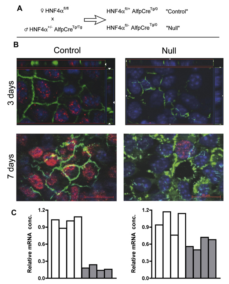
Fig. 1.
Short-term ex vivo culture of hepatic cells from e18.5 (embryonic day) HNF4α-null liver restores polarity and partially rescues expression of the E-cadherin gene. (A) Breeding scheme used to generate null (HNF4αfl/− AlfpCreTg/0) and littermate control (HNF4aαfl/+ AlfpCreTg/0). (B) Double immunofluorescence for tight junction protein 1 (TJP1/ZO1, green), and HNF4α (red) with nuclei visualized by DAPI (blue) staining, carried out at 3 and 7 days of culture. The “3 days” photomicrographs have bars to show vertical distribution of the fluorescence signals through the culture (z-stack). Immunostaining for HNF4α is intense in the control cells at 7 days, and a single stained nucleus has been captured for the null culture. (C) E-Cadherin transcripts, for control (open bars) and HNF4α-null (filled bars) e18.5 livers (left) and e18.5 three day cultures of hepatic cells (right).

