Figure 1.
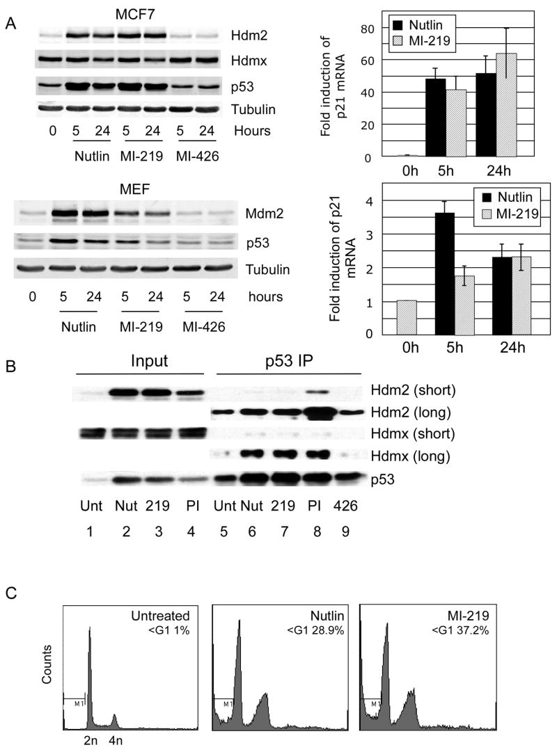
(A) MCF7 (upper panel) or MEFs (lower panel) were treated with Nutlin-3a, MI-219 or MI-426 (inactive control) all at 10uM for the indicated times and lysates evaluated for the indicated proteins. Quantitative qPCR of p21 mRNA abundance was used as a metric of p53 activation. (B) MCF7 were treated with Nutlin-3a, MI-219 or proteasome inhibitor MG132 (all 10uM) for 5h prior to immunoprecipitation with anti-p53 antibody FL393. Long and short refer to exposure times for Hdm2 and Hdmx. (C) SJSA were treated with Nutlin-3a or MI-219 for 36h and subjected to FACS analysis following propidium iodide staining.

