
Figure 5. Intracellular killing of P. gingivalis by AM.
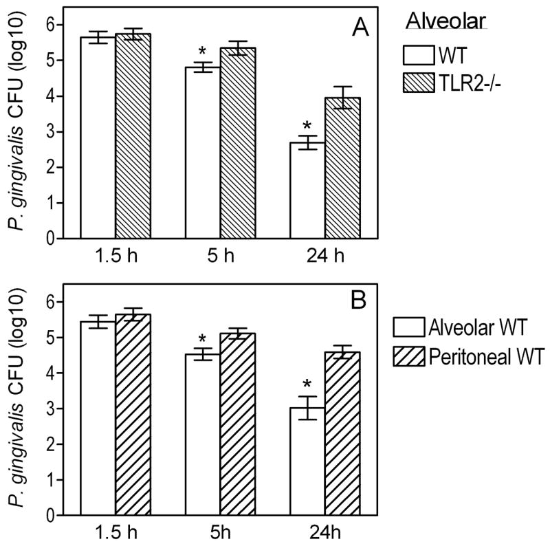
(A) Wild-type and TLR2−/− AM were incubated with P. gingivalis (MOI=10:1) for the indicated times. The persistence of viable internalized bacteria was determined using an antibiotic protection-based survival assay. (B) Similar procedures were followed to measure the intracellular killing of P. gingivalis by alveolar and peritoneal macrophages. Data are means ± SD (n = 5). Asterisks show significantly (p < 0.05) lower CFU levels (i.e., increased killing) in AM compared to their TLR2−/− counterparts (A) or to peritoneal macrophages (B).