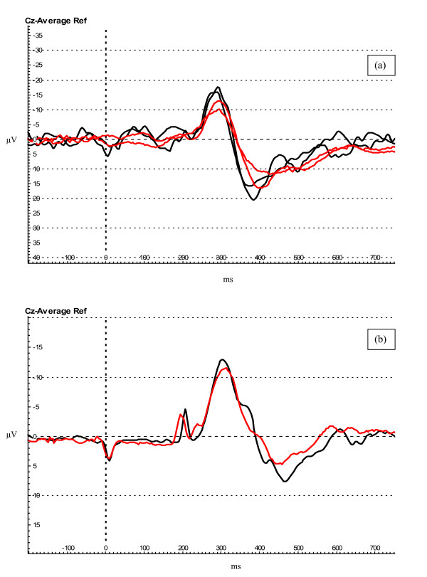
Figure 2.
Evoked potential waveforms. (a) Evoked potentials recorded during baseline (black traces) and fMRI (red traces) protocols in subject # 5. (b) Evoked potentials (grand average for all subjects) recorded at baseline (1 plus 2) (black trace) and during fMRI (1 plus 2) protocols (red trace). The deflections on the trace occurring at 0 and 250 ms are artefacts produced by the stimulator.

