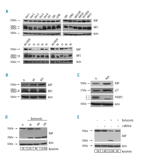
Figure 1.
XIAP and XAF-1 expression in myeloma cells. (A) XIAP expression was assessed by immunoblot analysis in HMCLs (top panel) and in a series of primary myeloma cells (P1–P9) (bottom panel). XAF-1 expression evaluated as indicated. Protein loading was controlled with anti-actin mAb. (B) XIAP and XAF-1 protein expression in the XG6 HMCL after culture in presence of IL6 (10 ng/mL corresponding to 385 nmol/mL) or IGF-1 (100 ng/mL corresponding to 13.1 μmol/mL) for 48h. (C) XIAP, p27 and p4EBP1 protein expression in untreated XG6 cells (Ct) or after rapamycin (Rapa) treatment (100 nM) for 24h. 3 isoforms of P4EBP41 were detected (a: hypophosphorylated isoform, b and g: Thr 37, 46- and 37, 46, 65, 70-phosphorylated isoforms respectively). (D) XIAP protein expression during bortezomib 10 nM treatment for 8, 16 and 24h. Specific apoptosis was evaluated in each condition by Apo2.7 immunostaining. (E) XIAP protein expression during bortezomib treatment in presence of the pancaspase inhibitor z-VADfmk (50 μM added one hour prior to bortezomib treatment) or not. Specific apoptosis was evaluated in each condition by Apo2.7 immunostaining.

