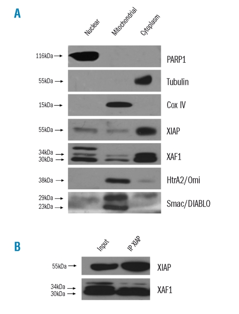
Figure 4.
XIAP and XAF-1 colocalized in the cytosolic fraction and were co-immunoprecipitated. (A) XIAP and XAF-1 localizations were evaluated by immunoblot analysis after subcellular fractionation of NCI-H929 cells. Enrichment of fractions was verified by expression of PARP1, COX IV and tubulin for nuclear, mitochondrial and cytoplasmic fraction respectively. (B) XIAP and XAF-1 co-immunoprecipitation in NCI-H929 cells. First lane (Input) corresponds to the cell lysate used for immunoprecipitation (IP) and second lane (IP XIAP) corresponds to the lysate obtained after IP.

