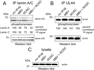
Figure 3. Orthophosphate labeling of lamin A/C during HCMV infection.
HCMV-infected cells (MOI = 3, 69 hours p.i.) were pulse labeled for 2 hours with [32P]-orthophosphate in the presence or absence of maribavir or roscovotine or both and then lysed. (A) Lamin A/C immunoprecipitates were prepared, resolved by SDS-PAGE, and transferred to a PVDF membrane. The membrane was exposed to a phosphorscreen (top panel, phosphorscreen) to detect 32P signal, and then probed with anti-lamin A/C antibodies to detect total lamin A/C in the immunoprecipitates (bottom panel, western blot). The signal from the phosphorscreen was quantified and the values for 32P incorporation into lamin A and lamin C are shown between the upper (phosphorscreen) and lower (western blot) panels. (B) HCMV UL44 immunoprecipitates were prepared from lysates of the same radiolabeled infected cells, resolved by SDS–PAGE and transferred to a PVDF membrane. The membrane was exposed to a phosphorscreen (top panel, phosphorscreen) to detect 32P signal, and then probed with anti-UL44 antibodies antibodies to detect total UL44 in the immunoprecipitates (bottom panel, western blot). The signal from the phosphorscreen was quantified and the values for 32P incorporation into UL44 are shown between the upper (phosphorscreen) and lower (western blot) panels. (C) The lysates used for immunoprecipitation in A were resolved by SDS-PAGE and transferred to a PVDF membrane that was probed with anti-actin antibodies. Abbreviations: IP, immunoprecipitate; MBV, maribavir; ROSC, roscovitine. The results show that transient inhibition of UL97 with maribavir reduces phosphorylation of lamin A/C by about 50%.

