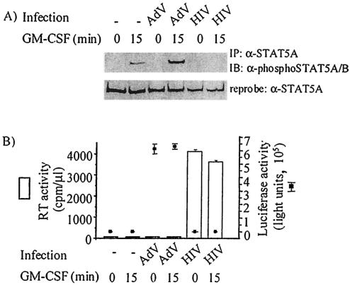
FIG. 4.
HIV-1, not adenoviral, infection of MDM inhibits GM-CSF-induced STAT5A tyrosine phosphorylation. Monocytes were cultured for 6 days prior to mock infection (−) or HIV-1Ba-L infection or for 10 days prior to infection using the ΔTARluc adenovirus (AdV) construct. After 7 days of HIV infection and 3 days of AdV infection, MDM were stimulated with 10 ng of GM-CSF per ml for 0 to 15 min and lysed. (A) Equivalent amounts of cell lysate were immunoprecipitated (IP) with an anti-STAT5A antibody (α-STAT5A), and the immunoblot (IB) was probed with an anti-phospho-STAT5A/B antibody (α-phosphoSTAT5A/B) and reprobed with an anti-STAT5A antibody. (B) Similar levels of HIV-1 infection in separate MDM cultures were demonstrated by RT activity in culture supernatants (in counts per minute per microliter of culture supernatant) taken prior to GM-CSF stimulation (left y axis; white bars). The level of AdV infection was assessed by lysing MDM which had been infected in parallel cultures to those used for immunoprecipitation and performing the luciferase assay on those lysates (right y axis; solid squares). The results shown are representative of MDM prepared from three donors.

