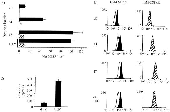
FIG. 5.
GM-CSFR α- and βc-chain surface expression is not inhibited by HIV-1 infection of MDM. Monocytes were cultured from day 0 in Teflon jars, and 4 days after isolation of monocytes, the cells were mock infected or infected with HIV-1Ba-L. (A and B) Surface expression of GM-CSFR α- and βc-chain was assessed in duplicate on the day of isolation (d0), 4 days after isolation (d4) and 3 days after HIV-1 infection (d7), using monoclonal antibody against the α-chain (4H1; 3 μg/ml; black bars and histograms) and βc-chain (1C1; 3 μg/ml; hatched bars and histograms) or an isotype-matched control IgG1 (MOPC 21; 2 μg/ml; white histograms). All samples were analyzed by flow cytometry. Fluorescence values were converted to molecules of equivalent soluble fluorochrome (MESF) units using a standard curve generated from calibration beads and the QuickCal program. Duplicate test samples were corrected for background fluorescence and averaged. The asterisks indicate that surface expression was not detected. (C) Seven days after HIV-1 infection, supernatants were removed from each MDM culture and assayed for HIV-1 RT activity with results expressed as counts per minute per microliter of culture supernatant. Results are representative of two experiments using MDM prepared from different donors.

