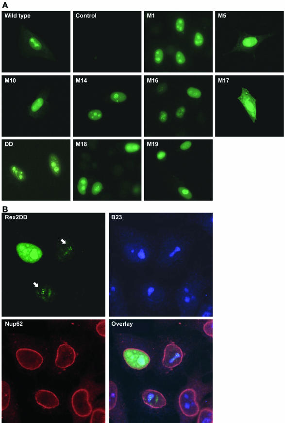
FIG.1.
Subcellular localization of Rex-2 mutants. The top set of panels (A) shows anti-Rex-2 immunofluorescence assays of HeLa-Tat cells that were transfected with wild-type rex or various rex mutant expression vectors. The bottom set of panels (B) shows laser scanning microscopy images of HeLa-Tat cells that were transfected with mutant DD and subjected to indirect immunofluorescence staining using antibodies against Rex-2, B23, and Nup62; the arrows point to DD-containing nucleolar speckles, and the pale blue-green and white areas in the overlay image indicate colocalization of DD with B23. Immunofluorescence assays were carried out as described in Materials and Methods.

