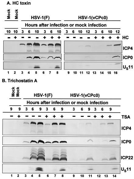
FIG. 5.
Temporal pattern of accumulation of selected wild-type HSV-1(F) and mutant HSV-1(vCPc0) proteins in RSC treated with H. carbonum (HC) toxin (A) and trichostatin A (B) 11 h before infection. (A) Replicate cultures of RSC in 25-cm2 flasks were either mock treated (−) or treated (+) with 70 ng of H. carbonum toxin per ml. At 11 h after treatment, cells were either mock infected (lanes 1 and 2) or infected with 5 PFU of wild-type HSV-1(F) per cell (lanes 3 to 8) or with 5 PFU of mutant HSV-1(vCPc0) per cell (lanes 9 to 16) in the absence (−) or presence (+) of 70 ng of H. carbonum toxin per ml. Cells were harvested at the indicated times after infection. Proteins were solubilized in disruption buffer, electrophoretically separated in 11% denaturing polyacrylamide gels, transferred to nitrocellulose sheets, blocked with 5% nonfat milk, and reacted with polyclonal antibody to ICP22 and monoclonal antibodies to ICP0, ICP4, and US11. (B) The experiment was repeated with 150 ng of trichostatin A (TSA) per ml, instead of H. carbonum toxin.

