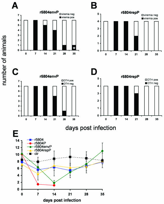
FIG. 8.
Cell-associated viremia, DTH responses, and in vitro lymphocyte proliferation activity of viruses with reassorted genes. (A and B) Cell-associated viremia detected after infection with r5804envP (A) and r5804repP (B). PBMCs were isolated weekly from 0.5 ml of blood and were cocultivated with VerodogSLAMcells. Cell-associated viremia was monitored by detection of syncytium formation between 1 and 3 days after the PBMC isolation. The white portions of the bars represent virus-negative animals, and the black portions represent virus-positive animals. (C and D) DTH responses of animals infected r5804envP (C) and r5804envP (D). Animals were immunized against tetanus prior to the experiment, and a positive DTH response was detected at the time of infection. The white portion of the columns represent animals with a positive DTH response, the black portions represent DTH-negative animals. (E) Comparison of in vitro proliferation activity of PBMC from animals infected the unaltered viruses r5804 (n = 4) and r5804P (n = 4) or the chimeric viruses r5804envP (n = 6) and r5804repP (n = 4). Methods were as described in the legend to Fig. 6. The color coding is as described for Fig. 7D. The proliferation activity of three noninfected control animals at each time point is indicated by black squares connected by an interrupted black line. Error bars are shown.

