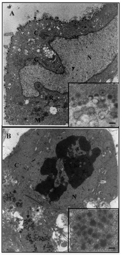
FIG.8.
TEM images of VZV-infected human DRG neurons and HFFs. (A) VZV-infected neuron at 2 days p.i. with an intact double nuclear membrane (NM), normal Golgi apparatus (G), mitochondrion (M), and endoplasmic reticulum (ER). The arrowhead and arrow indicate unenveloped nucleocapsids in the nucleus and enveloped virions on the cell surface, respectively. Magnification, ×6,500. (Inset) Higher magnification of enveloped VZV virions on the surface of the neuron. Bar, 200 nm. (B) VZV-infected HFFs at 2 days p.i. show morphological features of apoptosis, loss of double nuclear membrane, condensed chromatin (Chr), and irregular organelle morphology. An arrow indicates enveloped VZV virions in the cytoplasm. Magnification, ×6,500. (Inset) Higher magnification of enveloped VZV virions in the cytoplasm of the HFF. Bar, 200 nm.

