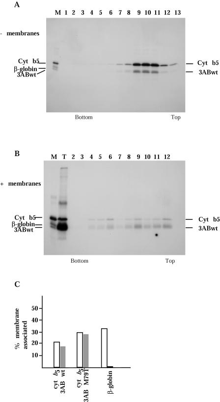
FIG. 5.
Sucrose density gradient analysis of 3AB proteins. (A and B) Translation reactions were programmed with FLAG epitope-tagged wild-type 3AB and cytochrome b5 in the absence (A) or presence (B) of canine pancreas microsomal membranes. Products were sedimented through discontinuous sucrose gradients, and fractions were collected and immunoprecipitated with anti-FLAG antibody before analysis on SDS-PAGE gels. A mixture of individually translated FLAG epitope-tagged wild-type 3AB, cytochrome b5, and β-globin proteins served asmarkers for the gel (lane M); lane T in panel B represents a sample of the translation reaction not subjected to gradient sedimentation. (C) Radioactivity in each fraction of the sucrose gradients containing proteins from translation reactions programmed with cytochrome b5 mRNA and either wild-type 3AB, 3AB-M79T, or β-globin mRNAs, in the presence of canine pancreas microsomal membranes, was quantitated by phosphorimager. The percentage of total radioactivity cosedimenting with membranes (fractions 2 to 6) was calculated for each protein and plotted.

