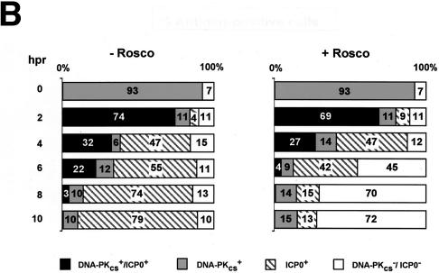
FIG. 6.
DNA-PKCS- and ICP0-specific immunofluorescence in WI-38 cells infected with KOS or 7134. (A) At 0, 4, and 8 h p.r. of the CHX block in the absence (−) and presence (+) of Rosco (R), infected cells were washed, fixed, permeabilized, and probed with primary and secondary antibodies to detect DNA-PKCS and ICP0 by immunofluorescence. Individual KOS-infected cells which stained positive for both DNA-PKCS and ICP0 (t = 4 h p.r. of the CHX block) are indicated by the arrows. (B) Percentages of DNA-PKCS+/ICP0+, DNA-PKCS+, ICP0+, and DNA-PKCS−/ICP0− staining in KOS-infected cells at 0, 2, 4, 6, 8, and 10 h p.r. of the CHX block in the absence (−) and presence (+) of Rosco. At least 200 cells were counted per time point per treatment group. The results obtained for mock- and 7134-infected cells at 0, 2, 4, 6, 8, and 10 h p.r. (data not shown) were indistinguishable from those for KOS-infected cells at 0 h p.r. of the CHX block.


