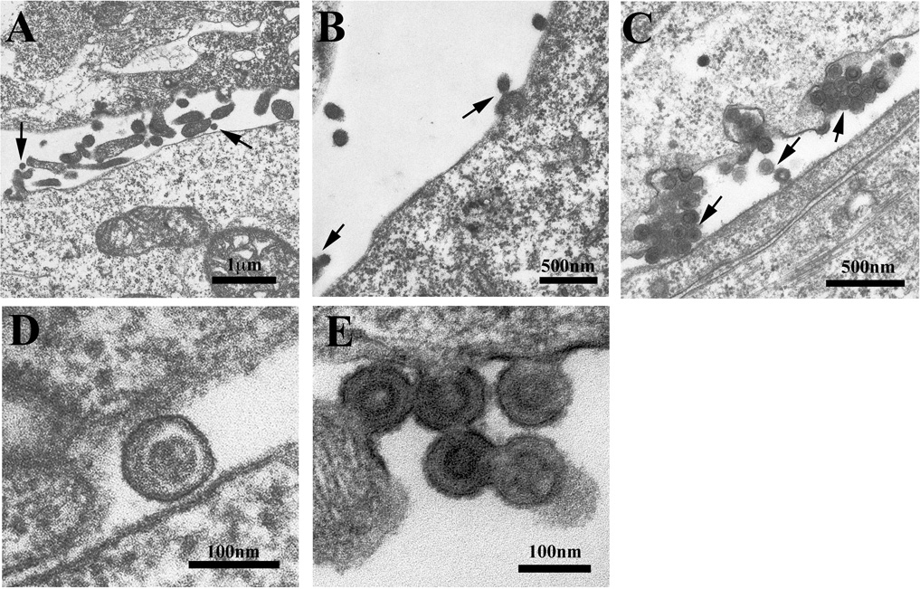
FIGURE 4.

Electron micrographs of 293T cells transfected with the following PERV provirus constructs: (A and D) PPPY-PFAP, (B and E) PPPD-PFAP and (C) PPPY-AAAA. Arrows indicate normal virion budding and phenotypes in (A), tethered viral particles at the cell membrane in (B) and dysmorphic grape-like clusters incompletely budded from the cell surface with tethered viral particles (middle arrow) in (C). Higher magnification images reveal aberrant PPPY-mutant cores compared to PPPY-PFAP (D and E). Micrographs are representative samples of multiple images obtained for each of the L domain mutant phenotypes.
