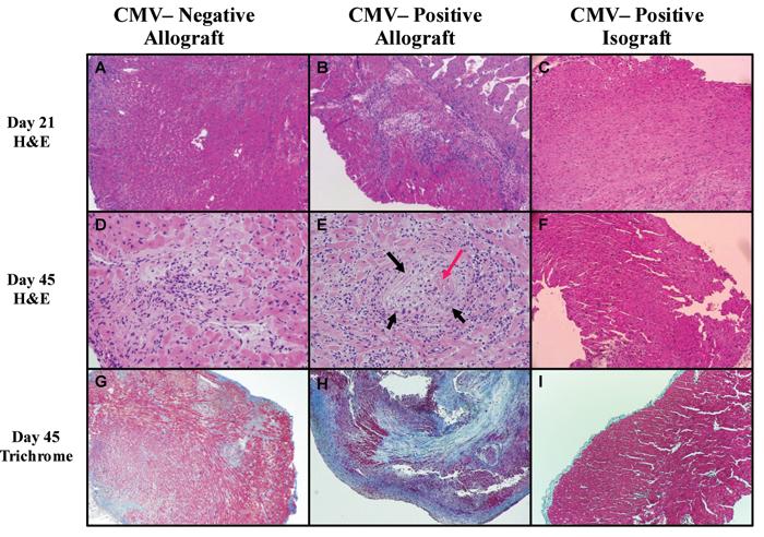
Figure 2. Pathologic changes in cardiac allografts of recipients latently infected with murine cytomegalovirus (MCMV).

Histology in cardiac allografts (BALB H2d) from MCMV-naïve C57BL/6 recipients compared to allografts and isografts from MCMV-positive C57BL/6 recipients 21 and 45 days post-transplant. A,D&C. Representative H&E (A&D) 21 and 45 days after transplant show typical cellular infiltration associated with this model, and trichrome sections (G) show mild patchy fibrosis in CMV-naïve recipient allografts. B&E. In contrast, CMV-positive recipient allografts show prominent cellular infiltration 21 days after transplant (B), which is more pronounced by day 45 (E) and is associated with vasculitis (black arrow) with fibrin (red arrow) deposition in lumen. H. Day 45 trichrome staining shows advanced fibrosis (blue staining). C,F,G. Isografts from CMV-positive recipients show bland myocardium, with very lile cellular infiltrate at either time point, and nominal fibrosis at day 45. Specimens were formalin fixed and embedded. Magnification = 40X, except for D & E 100X.
