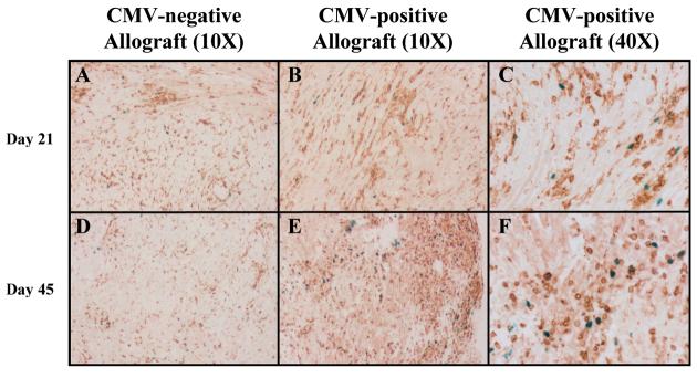
Figure 6. Foxp3 staining of cardiac allografts.
Immunohistochemical staining was performed on representative sections from CMV-positive and negative allografts 21 and 45 days after transplantation. A&D. Sections from CMV-negative allografts show numerous CD3+ cells (brown) with scattered Foxp3+ cells (blue). B&E. CMV-positive allografts show a similar staining pattern at day 21, but have more numerous Foxp3+ cells at day 45 after transplant. C&F. 40X views of CMV-positive grafts, clearly showing persistence of Foxp3+ T-cells (blue).

