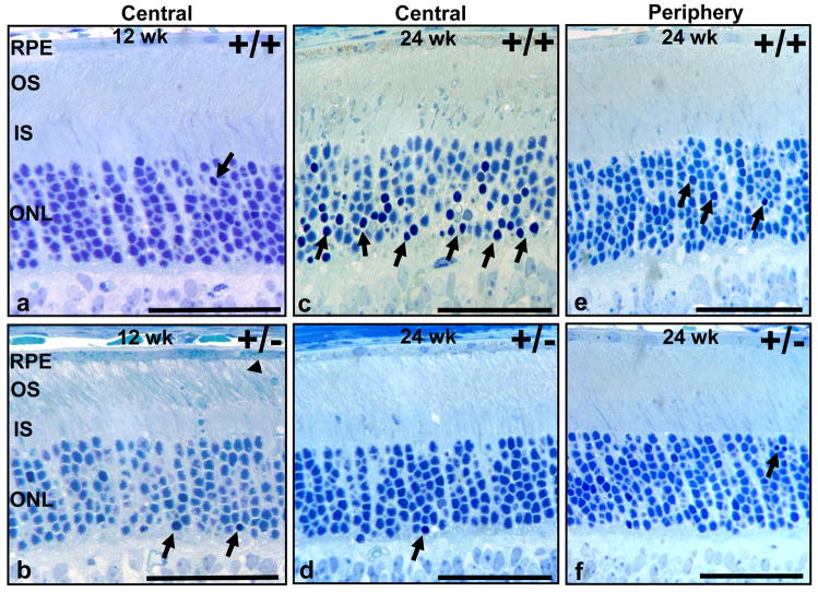
Figure 1.
Age-dependent light damage of photoreceptors is strongly reduced in RanBP2 haploinsufficient mice. Light photomicrographs of methylene blue-stained sections of central (a-d) and peripheral (e, f) regions of the retina of 12-(a, b) and 24-week old (c-f) RanBP2+/+ (a, c, e) and RanBP2+/− mice (b, d, f). There is a strong increase in pyknotic nuclei in 24-week old wild-type mice compared with 12-week old wild-type mice (arrows pointing to intense nuclei staining) in the central retina (a, c) that is accompanied by the disorganization of the outer nuclear layer. The periphery of the retina is spared largely from pyknosis (e). In contrast, no apparent differences in pyknosis are observed between 12- and 24-week old RanBP2+/− mice (b, d, f), but some vacuolization of the tip of the outer segments can be noted (arrowhead). Legend: RPE, retina pigment epithelium; OS, outer segment of rod photoreceptors; IS, inner segment of rod photoreceptors; ONL, outer nuclear layer (nuclei of photoreceptors). Scale bar, 40 μm.

