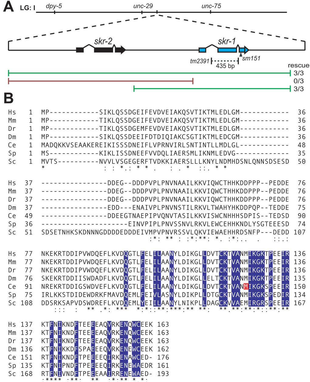
Figure 2.
(A) Schematic representation of the skr-2 and skr-1 locus of chromosome I (LG: I). The skr-1(sm151) lesion is indicated by an arrowhead and the skr-1(tm2391) deletion is indicated by a dashed line. Green lines represent PCR fragments that rescued skr-1(sm151) and the red lines represent PCR fragments that did not rescue. (B) Alignment was performed between Skp1-related proteins in human (Hs), mouse (Mm), zebrafish (Dr), fruitfly (Dm), worm (Ce), fission yeast (Sp), and budding yeast (Sc) with ClustalW2 (European Bioinformatics Institute of the European Molecular Biology Laboratory). (*) identical residues, (:) conserved residues, (.) semi-conserved residues. C. elegans SKR-1 M140 is indicated in red. F-box binding residues of human Skp1 are indicated in blue based on Schulmann et al. (2000).

