
FIG. 5.
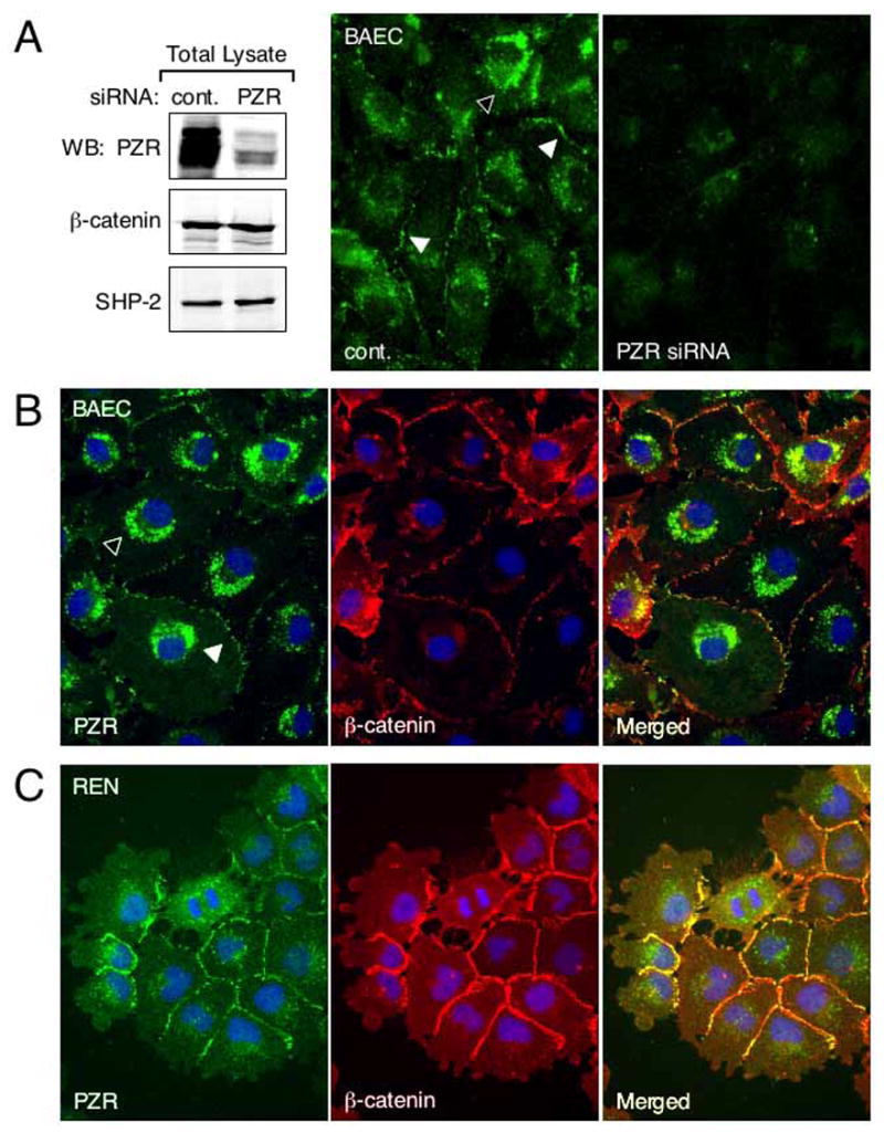
Subcellular localization of PZR. (A) BAECs were transfected with control or PZR siRNA duplex. Immunoblots show reduced PZR expression in siRNA treated cells. The expression of β-catenin and SHP-2 was not downregulated in these cells. When ECs transfected with control siRNA (cont.) were immunofluorescently stained with anti-PZR, cell contacts (white arrowheads) and some cytoplasmic granules (black arrowhead) were stained. This staining pattern was reduced in cells treated with PZR siRNA. (B) and (C) BAECs and REN cells were fixed and triple-stained with anti-PZR (green), anti-β-catenin (red), and DAPI for nuclear identification (blue). PZR was localized to cell-cell contacts (white arrowhead in BAECs) and intracellular granules (black arrowhead in BAECs). In merged images, yellow cell border staining is seen.