
FIGURE 5.
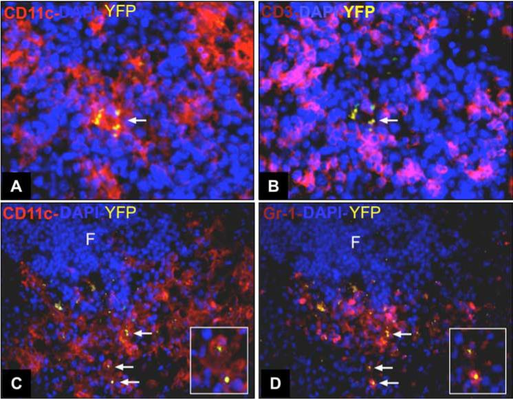
Infected cells in the T cell areas of the spleen coexpress CD11c and Gr-1. Mice were infected with RH-YFP tachyzoites and 4 days later spleens were collected for immunohistochemical staining. A, Four-color immunofluorescence staining of frozen sections shows CD11c+ cells (red) infected with RH-YFP (yellow). B, In the same section, staining for CD3 (pink) reveals T cells surrounding infected CD11c+ cells. Staining for CD11c (C) and Gr-1 (D) shows that most infected cells (arrows) coexpress CD11c (red) and Gr-1 (pink). Insets in C and D shows an expanded view of infected CD11c+Gr-1+ cells. Original magnification ×400 (A and B) and ×200 (C and D). This experiment was repeated twice with similar results.