
FIGURE 8.
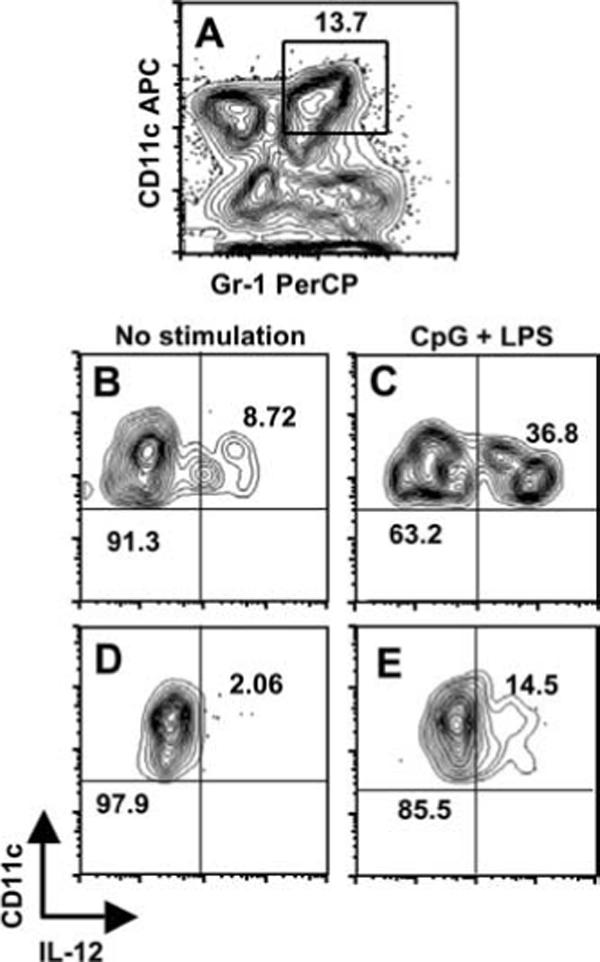
IL-12 production is defective in CD11c+Gr-1+ cells infected with T. gondii. Splenocytes were harvested 4 days postinfection with RH-YFP tachyzoites and enriched for CD11c+ cells using magnetic beads. A, CD11c and Gr-1 expression levels in the enriched DC population. The number indicates the percent of cells in the indicated rectangle. Enriched cells were cultured 24 h in medium alone, or medium with CpG oligodinucleotide (1 μg/ml) and LPS (100 ng/ml). The cells were stained for intracellular IL-12p40 and surface Gr-1 and CD11c. B, IL-12 expression by noninfected CD11c+Gr-1+ cells cultured in medium alone. C, IL-12 expression by parasite-negative CD11c+Gr-1+ cells after in vitro stimulation with LPS + CpG. D, IL-12p40 expression by YFP+ CD11c+Gr-1+ cells with no in vitro stimulation. E, Expression of IL-12p40 in infected CD11c+Gr-1+ cells after TLR ligand-stimulation. In B–D, the numbers indicate the percent of cells falling within the indicated quadrants. One representative of two independent experiments is shown.