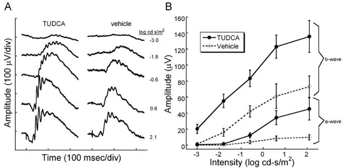
Figure 1.
Dark-adapted retinal function in rd10 mice after TUDCA treatments at P30. ERG recordings showing rod-dominated retinal function in P30 rd10 mice treated with TUDCA or vehicle. A) Representative ERG waves to a series of flash intensities from a TUDCA- or vehicle-treated mouse. At every light level, the TUDCA-treated mice had a larger response than the vehicle-treated rd10 mouse. The a-wave is much larger in the TUDCA-treated animals. B) Plot of average ERG amplitude (±SEM) for a- and b-waves from the two treatment groups. TUDCA (N=16) and vehicle (N=10). TUDCA-treated animals had significantly greater a- and b-wave amplitudes than the vehicle groups [Repeated measures ANOVA; a-wave: F(4, 88) = 2.66, p = 0.038; b-wave: F(4, 96) = 2.55 p = 0]. We conclude that TUDCA is efficacious in preventing the loss of ERG signals in response to a range of scotopic flashes.

