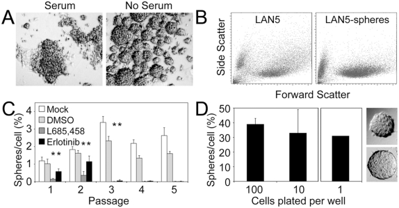
Figure 1. Neuroblastoma cells form tumorspheres in serum-free media.
(A) LA-N-5 neuroblastoma cells plated in serum supplemented media show adherent growth while those plated in serum-free media (with EGF and bFGF) formed non-adherent tumorspheres. (B) Dissociated bulk and tumorsphere cells were subjected to FACS analysis. Tumorsphere cells showed increased uniformity in complexity (low side scatter) compared to bulk cultured cells. (C) Tumorsphere formation over serial passage (plating at supra-clonal density) in neurosphere media alone or supplemented with a γ-secretase inhibitor (L685,458, 1 mM) or an EGFR inhibitor (erlotinib, 10 µM). The inhibitors were dissolved in DMSO, which was used as a negative control. (D) Plating of dissociated LA-N-5 tumorsphere cells at 100 or 10 cells per well showed consistent sphere-forming efficiency regardless of plating density; 30% of wells plated with a single cell contained a single sphere. Pictures show examples of clonally derived spheres.

