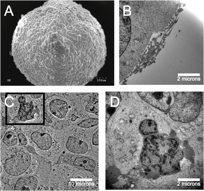
Figure 2. Ultrastructural characterization of neuroblastoma tumorspheres shows similarities to normal neurospheres.
(A) Scanning EM of neuroblastoma tumorsphere shows a smooth and uniform surface. (B) Microvilli-like structures shown on the tumorsphere surface by transmission EM in cross-section. (C) Transmission EM shows tight packing of tumor cells in the tumorsphere core. (D) Apoptotic tumorsphere cell shows nuclear blebbing, chromatin fragmentation and mitochondrial swelling.

