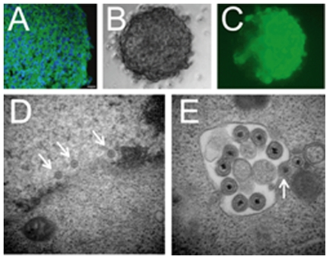
Figure 7. Neuroblastoma tumorspheres express nestin and are efficiently infected by oHSV.
(A) Immunohistochemistry on tumorsphere cryosection for nestin (green) and DAPI co-stain (blue). Scale bar = 10 microns. (B, C) A neuroblastoma tumorsphere infected with rQNestin34.5 was imaged at 48 hours post-infection by (B) phase-contrast and (C) fluorescent microscopy for GFP. (D, E) rQNestin34.5-infected neuroblastoma tumorsphere at 48 hours post-infection evaluated by transmission electron microscopy showing viral nucleocapsids in the nucleus (arrows), and mature HSV particles in the cytoplasm in the process of acquiring their envelopes (arrows).

