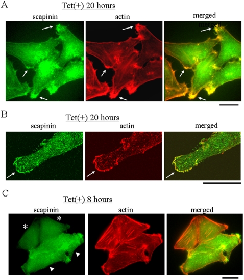
Figure 5. Distribution of scapinin and actin in Hela cells.
(A) Hela cells grown on glass coverslips were cultured in the presence (Tet+) or absence (Tet−) of 0.1 µg/ml tetracycline and were then fixed at 20 hours. After permeabilization, the distribution of scapinin and the actin cytoskeleton were visualized by staining with anti-scapinin antibody (green) and rhodamine-phalloidin (red), respectively. Scapinin and actin are colocalized (arrows). (B) Confocal microscopic observation. Hela cells were cultured in the presence of 0.1 µg/ml tetracycline for 20 hours and were then stained with anti-scapinin antibody (green) and rhodamine-phalloidin (red) as in (A). Scapinin and actin are colocalized (arrows). Bar: 20 µm. (C) Absence of scapinin in actin stress fibers. Hela cells grown on a glass coverslip were cultured in the presence of 0.1 µg/ml tetracycline and were then fixed at 8 hours. The distribution of scapinin and the actin cytoskeleton were visualized by staining with anti-scapinin antibody (green) and rhodamine-phalloidin (red), respectively as (A). There are four cells; two cells express low levels of scapinin (asterisks), and the other two cells express high levels of scapinin (arrow heads). Bar: 20 µm.

