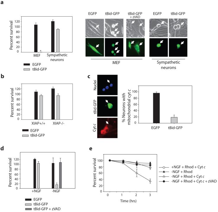
Figure 1. Endogenous cytochrome c release is incapable of inducing apoptosis in NGF-maintained sympathetic neurons.
a) Cultures of MEFs or sympathetic neurons were injected with tBid-GFP, or GFP plasmid. Cell survival was quantified by cell morphology and expressed as a percentage of alive and healthy green cells at 24 hrs compared to 5 hrs post-injection. *represents no MEF survival. Representative photographs of MEFs and neurons are shown; arrows point to injected cells. b) XIAP -/- sympathetic neurons or wildtype littermates were injected with EGFP or tBid-GFP, and cell survival was quantified as in (a). c) tBid-GFP was injected into sympathetic neurons, and allowed to express for 24 hrs. Cells were fixed, and the status of cytochrome c analyzed by immunofluorescence. Percentage of neurons with mitochondrial cytochrome c is quantified in XIAP -/- neurons. d) XIAP -/- sympathetic neurons were either maintained in NGF-containing media, or deprived of NGF for 8 hrs (with or without 50 μM zVAD), followed by injection with EGFP or tBid-GFP. Cell survival was quantified by cell morphology and expressed as a percentage of alive and healthy green cells at 16 hrs compared to 5 hrs post-injection. e) XIAP -/- sympathetic neurons were deprived of NGF (with or without 50 μM zVAD) for 12 hrs followed by injection with rhodamine alone, or cytochrome c (2.5 μg/μl) along with rhodamine to mark injected cells. Cell survival was assessed at various time points following injection. Error bars represent ±SEM of three independent experiments.

