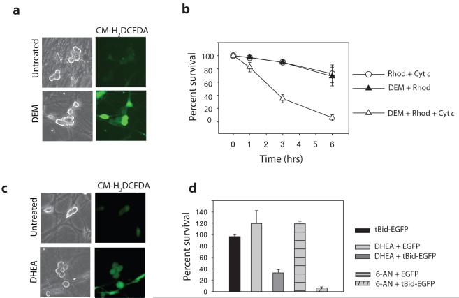
Figure 3. Role of the pentose phosphate shunt in cytochrome c-mediated apoptosis.
a) Average ROS levels in sympathetic neurons were observed by fluorescence intensity of the redox-sensitive dye CM-H2DCFDA following 30 min of GSH depletion with 0.1 mM DEM. b) XIAP -/- sympathetic neurons (NGF-maintained) were treated with 0.1 mM DEM for 30 min followed by injection of cytochrome c and rhodamine, or rhodamine dye alone. Cell survival was assessed at various timepoints. c) Average ROS levels were observed as in (a) following inhibition of the Pentose Phosphate Pathway with 200 μM DHEA for 24 hrs. d) XIAP -/- sympathetic neurons (NGF-maintained) were treated with 200 μM DHEA for 6 hrs, 0.1 mM 6-AN for 24 hrs, or left untreated, followed by injection with tBid-GFP or EGFP constructs. Cell survival was expressed as a percentage of healthy green cells at 16 hrs compared to 5 hrs post-injection. Error bars represent ±SEM of n≥3.

