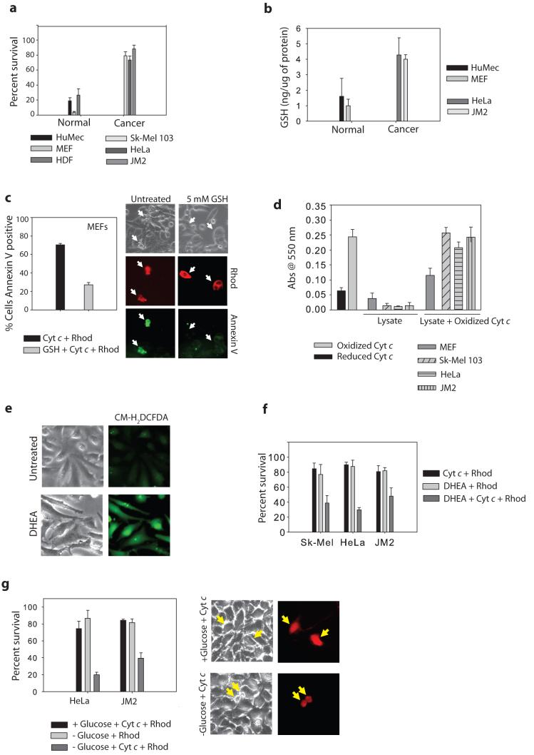
Figure 4. Glucose metabolism protects cancer cells from cytochrome c-mediated apoptosis.
a) Normal cells (Human Mammary Epithelial Cells-HuMECs, MEFs, Human Dermal Fibroblasts-HDFs) or cancer cell lines (Sk-Mel 103, HeLa, JM2) were injected with cytochrome c and Smac protein and cell survival was assessed after 30 minutes. b) Total glutathione (GSH) was measured in normal mitotic cells as well as cancer cell lines, and expressed as a concentration of GSH to total cellular protein. c) MEFs were treated with 5 mM GSH ethyl ester for 15 min, followed by injection with cytochrome c. After 1 hr, injected cells were assessed for Annexin V positivity. Photographs show representative Annexin V-FITC staining of cytochrome c injected cells (arrows). d) Exogenous cytochrome c (which is primarily oxidized) was added at a concentration of 10 μM to normal or cancer cell extract, and Abs550 was measured following a 15 min incubation. e) Average ROS levels in HeLa cells measured by fluorescence of CMH2DCFDA in the absence or presence of the pentose phosphate pathway inhibitor, DHEA (200 μM) for 6 hrs. f) The pentose phosphate pathway was inhibited in various cancer cell lines for 6 hrs by addition of 200 μM DHEA, followed by injection of cytochrome c and Smac protein. Cell survival was assessed at 3 hrs. g) JM2 and HeLa cells were deprived of glucose for 16 hrs followed by injection with cytochrome c and Smac, and assessed for survival at various time points. Images are representative of JM2 cells at 3 hrs following cytochrome c injection. Error bars represent ±SEM of n≥3.

