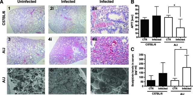
Figure 4.
A: Representative light micrograph (H&E-stained) (A1 to A4) and scanning electron micrograph (A5 and A6) of lung tissue sections of uninfected (left) or S. aureus-infected (middle and right) C57BL/6 and A/J mice. The lungs of infected C57BL/6 mice were unaffected (A2) and show similar alveolar architecture as lung tissue from uninfected mice (A1, A3). Widespread hemorrhages and massive erythrocyte infiltration of lung parenchyma and bronchioles were observed in lung sections of S. aureus-infected A/J mice (A4, A6). B: Activated partial thromboplastin time (aPTT) in plasma. C: Bradykinin release in serum of C57BL/6 (black bars) and A/J (white bars) mice at 24 hours after intravenous infection with 4 × 10⁷ cfu S. aureus. Bars represent the mean ± SD of five mice per group. *P < 0.05, by F-test. Scale bars: 100 μm (A5, A6i); 10 μm (A6ii). Original magnifications: × 40 (A1, A2i, A3, A4i); × 200 (A2ii, A4ii).

