Figure 6.
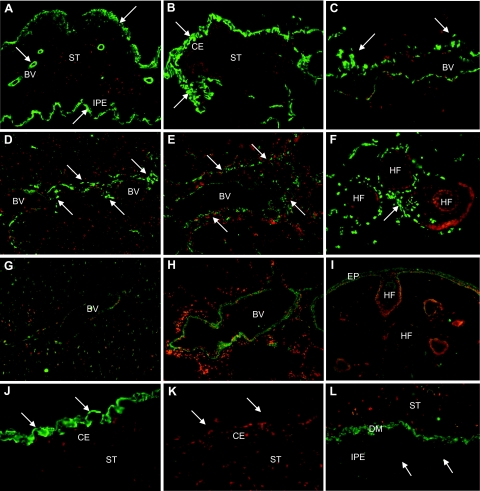
Immunolocalization of LOXL1 in PEX material deposits in various intra- and extraocular tissues of PEX eyes. Immunopositive PEX material deposits (arrows) are present in the iris stroma, periphery of iris vessels, and on the surface of the iridal pigment epithelium (A); on the surface of the ciliary epithelium (B); in the periphery of episcleral veins (C); in myocardial tissue (D); in lung tissue, particularly in the periphery of pulmonary blood vessels (E); and in the periphery of hair follicles in the skin (F). Corresponding control tissues show weak staining for LOXL1 in vessel walls of myocardial (G) and lung tissue (H) as well as in epidermal cells and root sheaths of hair follicles in normal skin (I). J and K: Pre-adsorption of LOXL1 antibodies with the corresponding peptide completely abolished immunopositivity of PEX deposits (arrows) on the surface of the ciliary epithelium. L: PEX material deposits (arrows) in the iris were negative for LOX. BV, blood vessel; CE, ciliary epithelium; DM, dilator muscle; EP, epidermis; HF, hair follicle; IPE, iris pigment epithelium; ST, stroma. Original magnifications, ×100.

