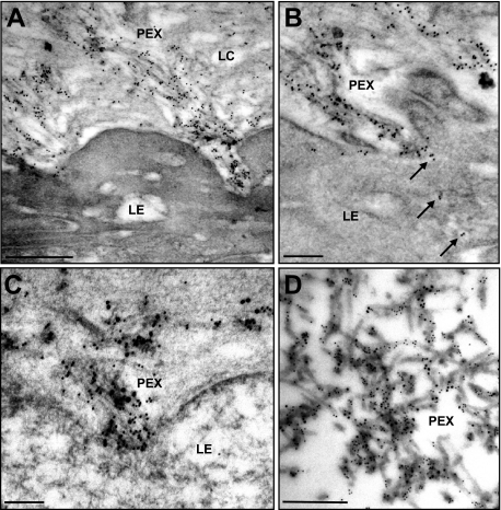
Figure 8.
Immunogold localization of LOXL1 on ocular PEX material. A: Immunopositive PEX fibers emerging from the pre-equatorial lens epithelium (LE) into the lens capsule (LC). B: Gold marker for LOXL1 is evident within cytoplasmic vesicles (arrows) of the lens epithelium (LE) and along PEX fibers on its surface. C: Co-localization of LOXL1 (10-nm gold particles) and fibrillin-1 (18-nm gold particles) along developing PEX fibers in close association to the surface of the lens epithelium (LE). D: Co-localization of LOXL1 (10-nm gold particles) and fibrillin-1 (18-nm gold particles) along mature PEX fibers on the surface of the lens capsule. Scale bars = 0.5 μm.

