Figure 1.
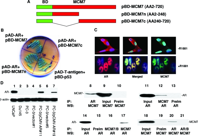
MCM7 interaction with AR. A: Schematic diagram of pBD-MCM7, pBD-MCM7n, and pBD-MCM7c. B: Growth in SD-4 high stringency agar plate and α-galactosidase activity of co-transfection isolates of yeast harboring pAD-AR+pBD-MCM7, pAD-AR+pBD-MCM7n, pAD-AR+pBD-MCM7c, and pAD-T-antigen+pBD-p53 (positive control). C: MCM7 and AR co-localization in nuclei of LNCaP cells induced with or without R1881. These cells were stained with antibodies against MCM7 (mouse monoclonal and FITC-conjugated donkey anti-mouse antibodies) and AR (rabbit polyclonal and rhodamine-conjugated donkey anti-rabbit antibodies). D: Expression of AR and co-immunoprecipitation of MCM7 and AR. Immunoblot analysis of AR from LNCaP (lane 1), DU145 (lane 2), and PC3 (lane 3), PC3 clone 1 transfected with pCMVscript (lane 4), PC3 clone 11 transfected with pCMV-AR (lane 5), PC3 clone 5 transfected with pCMVscript (lane 6), PC3 clone 19 transfected with pCMV-AR (lane 7). Co-immunoprecipitation was performed on protein extracts from LNCaP cells induced with R1881 (lanes 8 to 13). AR (lane 8) or MCM7 (lane 12) was immunoprecipitated and blotted with antibodies against MCM7 (lanes 8 to 10) or AR (lanes 11 to 13). Lysate input (lanes 9 and 11) was used as positive control for blotting, whereas pre-immune serum (PreIm) precipitation (lanes 10 and 13) was used as negative control for precipitation. Co-immunoprecipitation of AR and MCM7 from PC3 cells transfected with pCMV-AR (clone 11) was similarly performed using MCM7 (lane 17) or AR (lane 20) antibodies for immunoprecipitation, and blotted with AR (lanes 14 to 17) or MCM7 (lanes 18 to 21) antibodies. MCM7/B denotes the primary immunocomplexes of MCM7 incubation with plain Sepharose instead of protein G-conjugated Sepharose.

