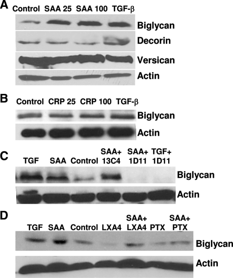
Figure 3.
SAA stimulates biglycan via TGF-β and the FPRL1 receptor. A: Cells were stimulated with SAA at 25 or 100 mg/L, or with TGF-β (2 ng/ml). B: Cells were stimulated with CRP at 25 or 100 mg/L, or with TGF-β (2 ng/ml). C: Cells were stimulated with TGF-β (2 ng/ml), SAA 100 mg/L alone or in combination with TGF-β neutralizing antibody 1D11 (10 μg/ml), or with irrelevant control antibody 13C4 (10 μg/ml). D: Cells were stimulated with TGF-β (2 ng/ml), SAA 100 mg/L alone or in combination with Lipoxin A4 (LXA4, 5μmol/L), or PTX (0.5 μg/ml) for 24 hours. Proteoglycan core protein synthesis was analyzed by Western blot. Lanes were loaded with an equal amount of protein, and blotted for biglycan, versican, decorin, and actin. Blots shown are representative of three independent experiments (A) or two independent experiments (B--D).

