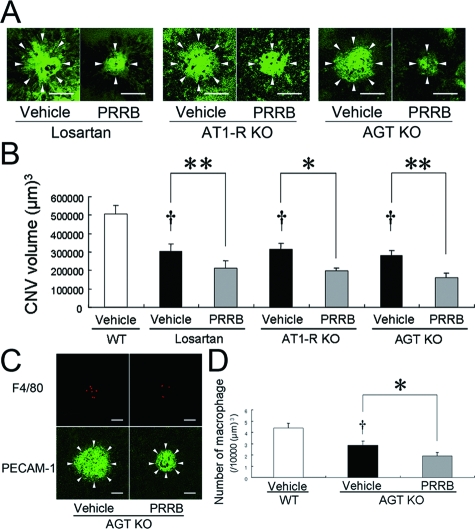
Figure 3.
RAS-independent (pro)renin receptor-mediated intracellular signaling contributes to CNV development and macrophage infiltration. The graph shows the choroidal flatmounts (A) and the CNV volume (B). PRRB treatment further induced a significant decrease in the CNV volume in losartan-treated, AT1-R-deficient and AGT-deficient mice. Arrowheads in (A) indicate lectin-stained CNV tissues (n = 23 to 40). Scale bars = 100 μm. F4/80-positive macrophages (C, top) and PECAM-1-stained CNV (arrowheads in C, bottom) were evaluated in AGT-deficient mice, and the volume-adjusted number of macrophages is shown in the graph (D). PRRB further caused significant suppression of macrophage infiltration (n = 14 to 17. †P < 0.01, **P < 0.01, *P < 0.05). Scale bars = 50 μm.

