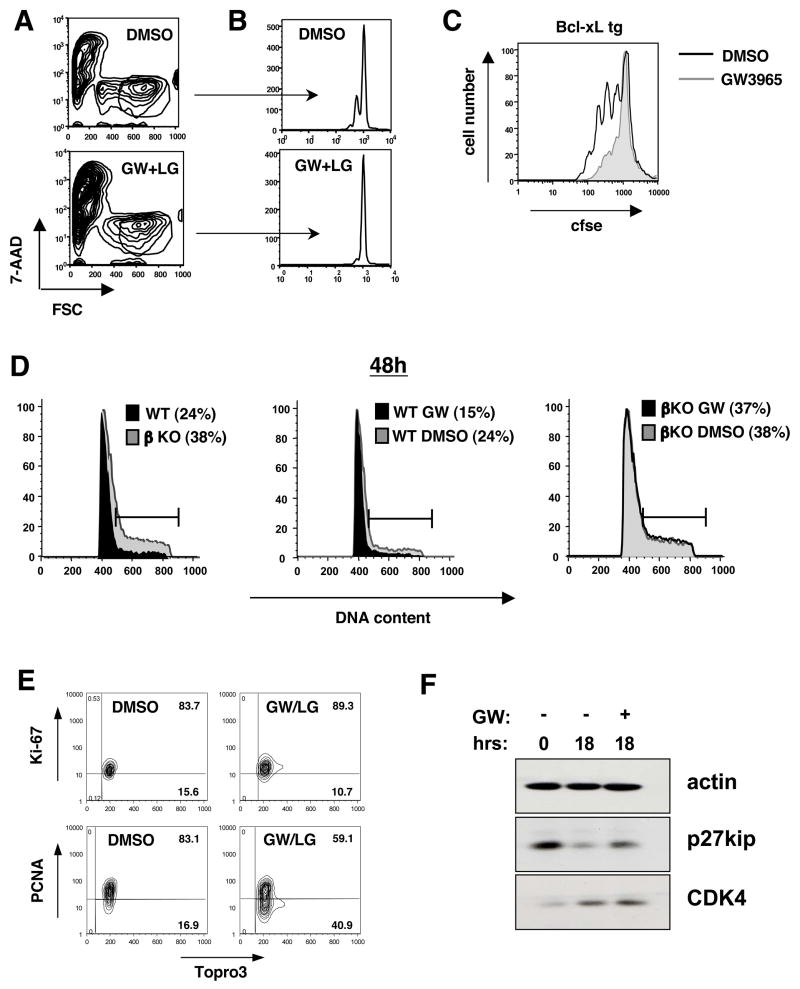
Figure 3. LXRβ signaling regulates cell cycle progression.
(A,B,C) CFSE dilution of WT and Bcl-xL tg T cells stimulated with pbCD3 (10μg/mL) for 36–96 h in the presence of GW3965 (2μM) and LG268 (100nM). Cells were stained with 7-AAD and analyzed by flow cytometry. (D) Cell cycle analysis of WT and LXRβ KO T cells stimulated with pbCD3 and GW3965 and LG268 as indicated. Cells were stained for DNA content with propidium iodide at 48h and analyzed by flow cytometry. (E,F) Cell cycle proteins of WT T cells stimulated with pbCD3 and GW3965 and LG268 as indicated. (E) Cells were permeabilized and stained for intracellular proliferation antigens Ki-67, PCNA and topro-3 for DNA content at 36h. (F) Whole cell lysates were collected and analyzed by Western blot for p27kip and CDK4 expression at 18h.

