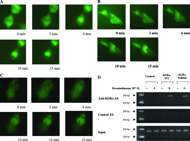
Figure 2.
Nuclear translocation of pF25GFP-hGRα (A) pF25GFP-hGRαD401H (B), and pF25GFP-hGRα in the presence of pRShGRαD401H (C) before and after exposure to dexamethasone. HeLa cells transiently expressing pF25GFP-hGRα or pF25GFP-hGRαD401H were exposed to the same concentration of dexamethasone (10−6 m). Images of the same cells were obtained at the indicated time points. There were no statistically significant differences between the wild-type (WT) hGRα and mutant receptor hGRαD401H in the time required for nuclear translocation after exposure to the ligand. D, ChIP assays performed on HCT-116 cells, in which the MMTV promoter was stably integrated into the chromatin. Both hGRα and hGRαD401H coprecipitated with MMTV glucocorticoid response elements in a ligand-dependent fashion, indicating that the mutant receptor hGRαD401H preserves its ability to bind to DNA. Ab, Antibody.

