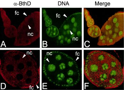
FIG. 3.
Localization of BthD in developing egg chambers. BthD antibodies (α-BthD) and DNA dye were used (as described in Materials and Methods) to visualize BthD expression (shown in red) (A and D) and DNA (shown in green) (B and E). (C and F) Merged images. (D to F) Higher-magnification images of the stage 8 egg chamber show that BthD is expressed throughout the cytoplasm of the follicle (fc) and nurse cells (nc). Magnification for panels A to C is ×100 and for panels D to F is ×200.

