Figure 2.
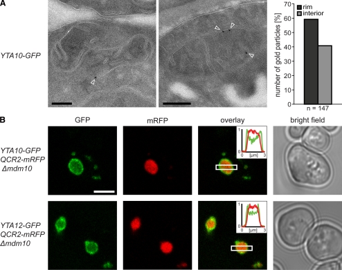
The m-AAA protease is preferentially localized in the IBM. (A) Localization of Yta10-GFP by quantitative immuno-EM in mitochondria of S. cerevisiae cells using GFP-specific antiserum. Left, representative images. The arrowheads point to gold particles. Right, quantification of the distribution of Yta10-GFP in the IBM (rim) and the CM (interior). (B) Live-cell fluorescence microscopy of Yta10-GFP or Yta12-GFP in cells exhibiting enlarged mitochondria due to a lack of Mdm10. Qcr2-mRFP was coexpressed to label the CM. Single confocal sections are displayed. Insets, normalized intensity profiles along the indicated area. Scale bars, (A) 200 nm; (B) 3 μm.

