Figure 3.
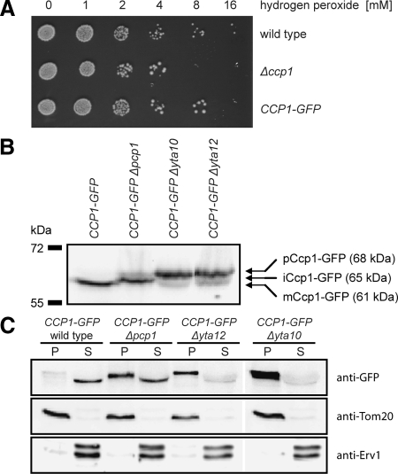
Ccp1-GFP is functional and processed correctly. (A) The growth phenotype of Δccp1 cells on elevated hydrogen peroxide concentrations is suppressed by the expression of Ccp1-GFP. Cells were grown to midlogarithmic growth phase and diluted in growth medium with the indicated hydrogen peroxide concentration. After 1 h at 37°C, 10 μl of the cell suspensions was spotted onto agar plates. (B) Analysis of Ccp1-GFP processing. Isolated mitochondria from wild-type, Δpcp1, Δyta10, or Δyta12 cells, each expressing Ccp1-GFP from the genomic locus, were analyzed by SDS-PAGE and analyzed by immunoblotting with GFP-specific antiserum to detect the fusion proteins. (C) Analysis of the membrane association of mCcp1, iCcp1, and pCcp1. Mitochondria isolated from cells expressing Ccp1-GFP and with the indicated genotypes were extracted with 200 mM sodium carbonate. The membrane pellet (P) and the soluble supernatant (S) were analyzed by SDS-PAGE and immunoblotting using GFP-specific antiserum. As control, antibodies against an integral protein of the outer membrane, Tom20, and a soluble protein of the IMS, Erv1, were utilized.

