Figure 4.
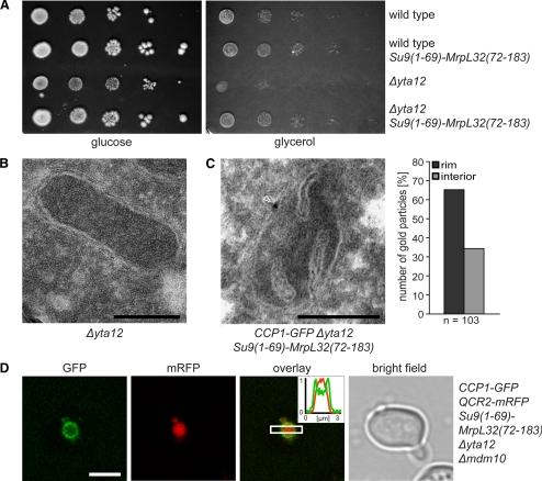
The precursor form of Ccp1 (pCcp1) is preferentially localized in the IBM. (A) Expression of the hybrid protein Su9(1-69)-MrpL32(72-183) suppresses the respiratory phenotype of Δyta12 cells. Tenfold serial dilutions of logarithmically growing cultures were spotted onto plates containing glucose or glycerol as sole carbon sources and incubated for 6 d at 30°C. (B) Electron micrograph showing that the mitochondria of Δyta12 cells exhibit a strongly reduced number of cristae. (C) Left, exemplary EM image of a mitochondrion of a cell lacking Yta12, but expressing the hybrid protein Su9(1-69)-MrpL32(72-183). The mitochondrion was immunogold-labeled for Ccp1-GFP; the arrowhead points to a gold particle. Right, quantification of the distribution of pCcp1-GFP in the IBM (rim) and the CM (interior). (D) Live-cell fluorescence microscopy of Ccp1-GFP coexpressed with the CM-marker Qcr2-mRFP in Δyta12 cells. To enable the light microscopic analysis, the mitochondria were enlarged by deleting MDM10. Single confocal sections are displayed. Scale bars, (B and C) 200 nm; (D) 3 μm.

