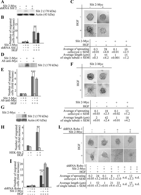
Figure 3.
Slit-2 inhibits HGF-dependent cell migration and tubulogenesis in MDA-MB-435 cells. (A) Anti-Myc Western blot expression of Slit-2-Myc in MDA-MB-435 cells. Cotransduction with the SLIT-2-specific shRNA B abrogates the expression of Slit-2-Myc. (B) HGF-dependent anisotropic migration in cells transduced with an empty vector, a Slit-2-Myc vector, or cotransduced with Slit-2-Myc together with the SLIT-2-specific shRNA-B. Cells were either left untreated or stimulated with HGF for 12 h. Data are the means ± SD (error bars) of three independent experiments performed in duplicate. **p < 0.01 at 12 h. (C) Tubulogenesis assay in cells expressing the empty (−) or the Slit-2-Myc (+) vector, in the presence of vehicle (−) or HGF (+). The micrographs show representative 10-d old colonies. Bar, 20 μm. Morphometric analysis is in the bottom panel. (D) Anti-Myc Western blot of conditioned media obtained from CHO cells infected with a control empty virus (−) or with a virus encoding for Slit-2-Myc (+). Immunodepletion of Slit-2 from the culture supernatants using anti-Myc antibodies is also shown. (E) Anisotropic migration of MDA-MB-435 cells in the presence of control supernatant, Slit-2–containing supernatant, or Slit-2–depleted supernatant. This analysis was performed after 12 h of HGF treatment. Data are the means ± SD (error bars) of three independent experiments performed in duplicate. ***p < 0.001. (F) Tubulogenesis assay in MDA-MB-435 cells in the presence of Slit-2–immunodepleted (−) or Slit-2–containing (+) medium, with or without HGF. The micrographs show representative 4-d-old colonies. Bar, 20 μm. Morphometric analysis is in the bottom panel. (G) Western blot expression of Slit 2-Myc in HEK cells. (H) Anisotropic migration of MDA-MB-435 cells cocultured with HEK cells expressing or not expressing Slit-2-Myc. Cell migration was quantitated after 12 h of HGF treatment. Data are the means ± SD (error bars) of two independent experiments performed in duplicate. **p < 0.01. (I) Anisotropic migration of MDA-MB-435 cells expressing a scrambled shRNA (−) or the ROBO-1–specific shRNA B (+) in the presence of Slit-2–containing (+) or Slit-2–depleted supernatant (−). This analysis was performed after 12 h of HGF treatment. Data are the means ± SD (error bars) of three independent experiments, performed in duplicate. ***p < 0.001 and *p < 0.05. (L) Tubulogenesis assay in MDA-MB-435 cells expressing a scrambled shRNA (−) or the ROBO-1–specific shRNA B (+) in the presence of Slit-2–containing (+) or Slit-2–depleted supernatant (−), with or without HGF. The micrographs show representative 10-d-old colonies. Bar, 20 μm. Morphometric analysis is in the bottom panel. n.d., not determined.

